3D X-Ray Microscopy for Material Science

Bruker’s portfolio of 3D X-ray microscopes offers turnkey solutions for non-destructive 3D imaging for a wide variety of industrial and scientific applications

This includes defect detection in casting, machining and additive manufacturing, inspection of complex electro-mechanical assemblies, pharmaceutical packaging, advanced medical tools, porosity and grain size analysis in geological samples, and in-situ microscopy.

SKYSCAN 1272

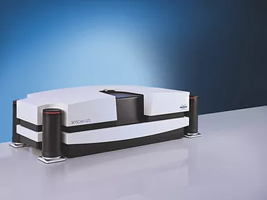
Our SKYSCAN 1272 can nondestructively visualize up to 209 Megapixel (14450x14450 pixels) virtual slices through objects, more than 2600 such slices after a single scan using a 16-megapixel camera in triple offset mode. Thanks to phase-contrast enhancement, object details as small as 0.35um can be detected.

The ability to move both the sample and large-format CCD camera as close as possible to the source substantially increases the measured intensity. That is why SKYSCAN 1272 scans up to 5 times faster compared to conventional systems with fixed camera position.

The SKYSCAN 1272 offers automatic selection of parameters with Genius-Mode. Magnification, energy, filter, exposure time and background corrections can all be optimized automatically with a single click.

Large format imaging is supported by multithreaded GPU-accelerated 3D-reconstruction and realistic visualization by surface and volume rendering. For high-speed requirements, InstaRecon®, the world’s fastest CT reconstruction software, accelerates 3D imaging up to 100 times compared to traditional algorithms.

SKYSCAN 1273

The SKYSCAN 1273 is Bruker’s latest benchtop 3D X-ray microscope based on micro computed tomography (Micro-CT) technology. Samples with up to 500 mm length, 300 mm diameter, and a maximum weight of 20 kg can be accommodated, a new standard for non-destructive testing (NDT) with benchtop instruments. Best-in-class components turn the SKYSCAN 1273 into a real power pack. The combination of a high-energy X-ray source running at high power and a large format flat-panel detector with ultimate sensitivity and speed provides excellent image quality in just a few seconds.

The comprehensive software for straightforward data collection, advanced image analysis, and powerful visualization makes the SKYSCAN 1273 an easy to use 3D X-ray microscope.

The SKYSCAN 1273 benchtop 3D X-ray microscope requires minimum lab space, is easy to start running, and virtually maintenance-free. Consequently, the SKYSCAN 1273 offers high system uptime with low cost of ownership.

SKYSCAN 1275

The SkyScan1275 is specially designed for fast scanning using new advances in the technology of X-ray sources and efficient flat-panel detectors. Shortening the distance between source and detector and very fast camera readout opens the possibility for reducing scan time down to a few minutes without compromising image quality. Current developments in fast reconstruction accelerated by graphics cards give an additional gain in performance and speed.

Fast realistic visualization of results by volume rendering enables fabulous imaging of internal object’s microstructure with power to reveal all internal details by virtual cut or virtual flight around and inside the objects. Fast scanning with high quality results is crucial for scientific research and industrial applications such as quality control or production process monitoring.

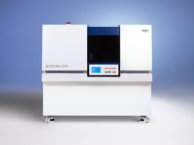
SKYSCAN 2214

The new MULTISCALE X-ray nanotomograph SKYSCAN 2214 covers the widest range of object sizes and spatial resolutions in one single instrument. It opens unique possibilities for 3D imaging and exact modeling of geological materials in oil and gas exploration, composite materials, Li batteries, fuel cells, electronic assemblies, as well as ex-vivo preclinical applications like in lung imaging or tumour vascularization.

The instrument allows scanning and 3D non-destructive reconstruction of internal microstructure of objects as large as >300mm in diameter as well submicron resolution for small samples. The world’s fastest hierarchical 3D reconstruction software speeds-up imaging object’s internal microstructure 10-100 times compare to traditionally used algorithms or GPU-accelerated reconstruction.

The system contains "open type" [pumped] X-Ray source with < 0.5 microns spot size and diamond window. It can be equipped with one to four X-Ray detectors: flat-panel for large objects, 11Mp cooled CCD with wide field of view, 11Mp cooled CCD with medium field of view, 8Mp cooled CCD for highest spatial resolution. Automatically variable acquisition geometry and phase-contrast enhancement allow obtaining best possible quality in relatively short scanning time.